1.Introduction
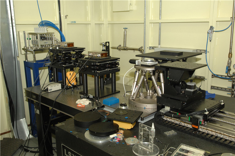
Since May 6, 2009, SSRF X-ray Imaging and Biomedical Application Beamline (BL13W1) has been put into operation and formally opened to users. BL13W1 is composed of a 1.9 Tesla wiggler, 6 set of filters for high heat load reducing, and double crystal monochromator cooled with liquid nitrogen. The monochromator can work well at the photon energy range of 8-72.5keV. Up to now, the filters and the monochromator could work well at 200mA, 3.5GeV and 17mm wiggler gap. At the end station, three sets of digital X-ray detectors are employed with the pixel size range from 0.37 to 13 micrometers. Imaging methods of microscopic computer tomography (micro-CT), in-line phase contrast imaging (IL-PCI) and X-ray fluorescence imaging could be provided for scientific researches on high Z and low Z samples in field of biomedical, material, archaeology, environmental science and so on. Spatial resolution of 0.8μm and time resolution of 1ms could be achieved. For X-ray fluorescence imaging, the minimal beam size is 30μm.
2.Scientific case
Soft tissues and low Z materials
Low dose, Nondestructive, High resolution, Dynamic and 3-dimensional X-ray Imaging for the inner microstructure
Paleontology, archaeology and geology
Nondestructive, High resolution, 3-dimensional X-ray Imaging for the inner microstructure
3.Imaging techniques
Absorption-contrast Imaging: Dynamic Imaging; Microscopic Computer Tomography
In-line Phase-contrast Imaging: Dynamic Imaging; Microscopic Computer Tomography
X-Ray Fluorescence Imaging: 2D scanning; Computer Tomography
4.Beamline Specifications
Unfocused monochromatic beam
Photon energy range: 8-72.5keV
Energy resolution (DE/E): <5×10⁻³
Beam size: 45mm (H) ×5mm (V) @34m@20keV
Flux output: 5×10¹⁰ phs/s/mm2 @ 20keV @ Si (111)
2×10⁸ phs/s/mm2 @ 70keV @ Si (311)