- Facilities at a Glance
- All Facilities
- Material
- Earth System and Environment
- Engineering Technology
- Space and Astronomy
- Particle and Nuclear Physics
- Energy
- Biology

- 1
- 2
- 3

Shanghai Institute of Applied Physics,CAS
Contact: Li Honghong
Phone: +86 021-20302750
Email: ssrf-user@sinap.ac.cn
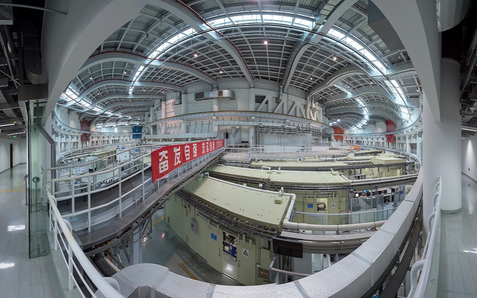
Shanghai Synchrotron Radiation Facility (SSRF)
Create Proposal Shanghai Synchrotron Radiation Facility (SSRF) is one of the advanced third generation light sources in the world, and it supports and pushes the cutting-edge scientific research and the innovation in China.Since commencing operation in May 2009 the SSRF has provided bright x-ray beams to more than 10000 users from both Chinese and international universities, institutes, hospitals and high-tech companies.
The facility has supported multiple scientific researches and industrial development, including biology, physics, material science,chemistry, environmental science, archeology, biomedical applications, medicine and drug development, etc.Over 1900 publications have been generated based on the experiments conducted at the SSRF.
The SSRF is also actively involved in the training and education of our next generation of scientists and engineers.The SSRF is committed to upgrading its capacity and strengthening its contribution to the cutting edge of modern science.
Equipment
-
Ambient pressure X-ray Photoneltron spectrascopy
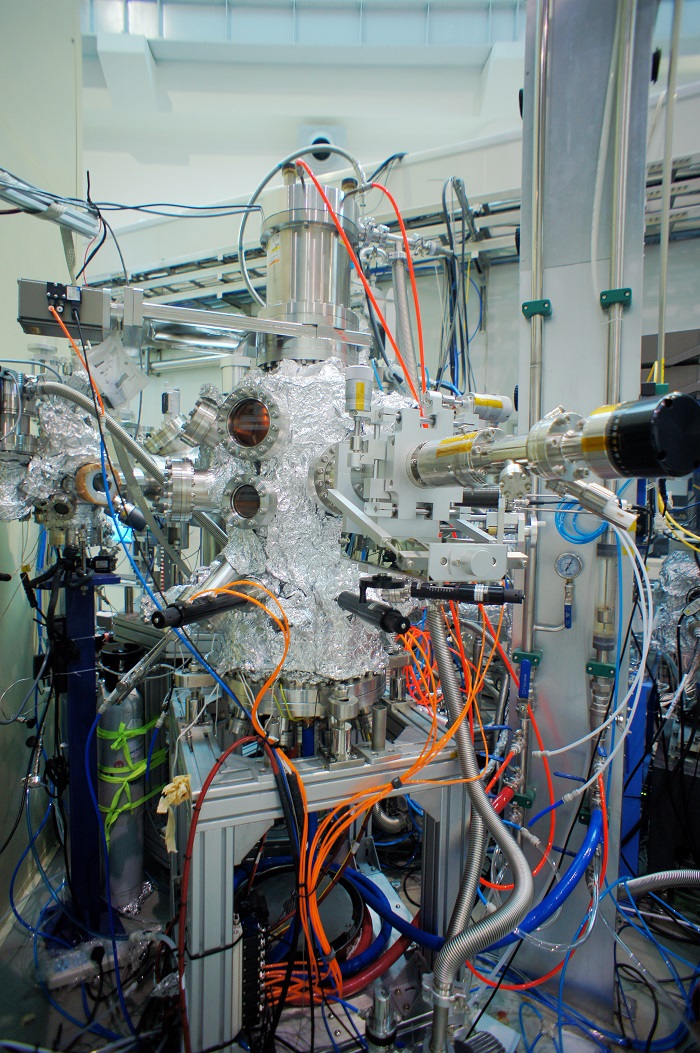
A new ambient pressure photoelectron spectroscopy (APPES) instrument has been installed at beamline 02B of Shanghai Synchrotron Radiation (SSRF). BL02B is a bending magnet beamline and has the photon energy ranging from 40 to 2000 eV, which has two branches. The APXPS system was equiped with an analyser of Scienta Omicron HIPP-3, which could be used in spatial mode. The new APPES instrument can measure samples at near ambient pressure up to 20 mbar. The new system owns the ability of spatia resolution up to 7.5 µm on spatial mode. Also, the APXPS system is collected with a multifunctional preparation chamber, which could be used for sample growth (evaporator), sample cleaning (Argon sputtering gun) and sample charaterization (LEED system).
-
Photon in Photon out End-station
BL02B is a bending magnet beamline and has the photon energy ranging from 40 to 2000eV, which has two branches of APPES and PIPOS endstation.
-
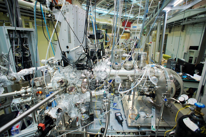
High-resolution angle resolved photoemission spectroscopy
The high-resolution ARPES experimental station (BL03U) uses the high-resolution, high-brightness vacuum ultraviolet beam of Shanghai Synchrotron Radiation Facitity (one of the best performing synchrotron radiations in the world) and tunable deep-UV solid-state laser source, and the independently developed internationally advanced sub femtosecond ultrashort second light source, overcomes the limitations of traditional electronic structure research in detecting depth, vertical momentum resolution and time resolution, which greatly improves the research capability and efficiency of the platform. Moreover, the platform integrates ARPES,MBE and STM, which makes up for the shortage of traditional electronic structure research in samples and improves the power of material control. Energy resolution of the BL03U platform: ~1 meV (photoemission spectroscopy), momentum resolution: ~0.003 Å-1, time resolution: ~10-16 s, which is at the international advanced level.
-

BL08U1-A Soft X-ray Spectromicroscopy Beamline
X-ray spectromicroscopy is a technique for high brilliant tunable synchrotron radiation, which could combine the two important functions such as the sub-50 nm resolving ability in space determined by the zone plate, and the chemical distinguishing ability determined by the beamline monochromator via NEXAFS spectroscopy. The photon energy of BL08U1-A was range from 250 to 2000 eV, covering the K-edge of C, N, O, F, Na, Mg, Al, Si, and L edges of Cl, K, Ca, Fe, Cu, Zn. The study of these elements is very important in biological science, environmental science, polymer, and material science. In addition, the light source was finally decided by an elliptical polarized undulator, which would add more opportunities to study x-ray-polarization-dependent materials.
-
BL08U1-B Soft X-ray Interference Lithography (XIL) Beamline
Soft-X ray interference lithography (XIL) is a newly developed technique for production of periodic nano-structures with resolution below 100 nm. The technique is based on coherent radiation obtained from undulators at synchrotron radiation (85-150 eV). Because of its small wavelength (typical value: 13.4 nm) and practical absence of the proximity effect, high density resolution lines/dots with high density can be afforded. The throughput of this parallel exposing method is much higher than that of the serial electron-beam lithography. Interference schemes based on diffraction (gratings) optics have been constructed at BL08U1-B beamline in SSRF. Both one-dimensional and two-dimensional patterns such as arrays of dots have been achieved. XIL is used in a growing number of applications; examples include fabrication of self-assembly templates, magnetic nano-dot arrays and nano-optical components.
-

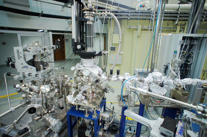
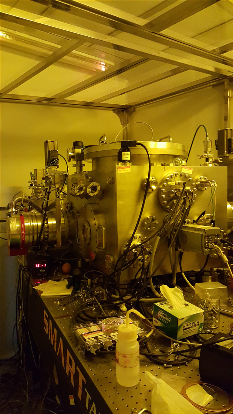
BL09U X-ray Photoemission Spectroscopy and Microscopy Beamline
1. Introduction
The X-ray Photoemission Spectroscopy and MicroscopyBeamline(Dreamline) provides a state-of-the-art experimental set-up to study the electronic band structure of novel complex materials and the surfaces and interfaces of solid-state materialsby using Angle-Resolved Photoelectron Spectroscopies (ARPES) and Photoemission Electron Microscopies (PEEM). The beamline was built with two APPLE-II type undulators(LEID and HEID) and produces a broad energy range from 20 to 2000 eV with high resolution, variable polarization, and high flux, as well as the minimized higher orders harmonics for the low energy photons. Due to this unique Duo-undulator technique, Dreamline provides an ideal platform for investigating the materials that has distinctproperties from the bulk to the surface.
2. Beamline Layout
Beamlie layout
Optics
3.Techniques
ARPES End-station
• UVU ARPES
• Soft X-rayARPES
• Resonant X-ray ARPES
• MBE
XPEEM End-station
• Bright & dark field LEEM
• MEM
• UV-PEEM
• X-ray PEEM
• LEED
• Spectroscopy
4. Endstations
ARPES End-station
The end-station isequippedwith a DA30L analyzer with the updated deflection mode capable for taking Fermi surface with high efficiency, and a 6-axis cryogenic manipulatorCarving®providing 3 translation and 3 rotation degree of freedom.This very solid and stable manipulator enables precise sample manipulating controlled by a sophisticateLabview-based software. The endstation also contains a load-lock with 6 parking position available, a transfer chamber providingAr-sputtering, a preparation chamber equipped with a LEED and a K-cell, and a magnetic shielded main acquisition chamber. Besides, the endstation has a MBE, which can be used for in-situ growing and photoemission studies forFe(TeSe) thin films.
The specification of the station is listed as below:
Eoptimal = 5 meV @ 20 eV, 21 meV @ 400 eV
Base temperature Tbase= 12 K (Carving) / 7 K (4-axis Manipulator)
Samples preparation: Ar-sputtering, annealing up to 1000℃,K-cell evaporators
In-situ Fe(TeSe) growth and characterization (MBE )
XPEEM End-station
The instrument allows to image samples using the photoelectric effect with very high spatial resolution, chemical and magnetic sensitivity. With an energy analyzer the excited photoelectrons can be energy-selected. In addition to illumination by X-rays, illumination by low energy electrons is possible. In this low energy electron microscopy (LEEM) mode additional contrast mechanisms are available.
The special/energy resolution data:
LEEM/PEEM: Field of view: 0.65-100um
Typical lateral resolution:
LEEM imaging: ~ 3nm
PEEM imaging: UV lamp: ~6nm
X ray: ~ 20nm
Energy resolution: ~0.15eV
Ion gun: sputter and clean sample surface
Femtosecond laser: 800nm and 400nm wavelength
Mercury lamp: photon 4. 9eV
Gas: O2, Ar, etc.
K-cell and E-beam evaporators: in situ growth and observation
5. Support Facilities
Glove Box: AUnilab Pro SP(1250/1000) glove box is available for preparing and storing air-sensitive samples
6. Beamline Specifications
Energy range 20-200 eV(LEID),200-2000 eV(HEID)
Resolving power 35000 @ 867 eV
Polarization Linear Horizontal, Linear Vertical,Circular Left/Right
Flux on sample 3.5×10¹¹ phs/s/0.01% BW @ 800eV
Beam spot 20 m×30 m
7. Applications
8. Contact
Yaobo Huang
huangyaobo@sinap.ac.cn
+86(0) 21 3393 3224 -

BL13W1 X-ray Imaging and Biomedical Applications Beamline
1. Introduction
Since May 6, 2009, SSRF X-ray Imaging and Biomedical Application Beamline (BL13W1) has been put into operation and formally opened to users. BL13W1 is composed of a 1.9 Tesla wiggler, 6 set of filters for high heat load reducing, and double crystal monochromator cooled with liquid nitrogen. The monochromator can work well at the photon energy range of 8-72.5keV. Up to now, the filters and the monochromator could work well at 200mA, 3.5GeV and 17mm wiggler gap. At the end station, three sets of digital X-ray detectors are employed with the pixel size range from 0.37 to 13 micrometers. Imaging methods of microscopic computer tomography (micro-CT), in-line phase contrast imaging (IL-PCI) and X-ray fluorescence imaging could be provided for scientific researches on high Z and low Z samples in field of biomedical, material, archaeology, environmental science and so on. Spatial resolution of 0.8μm and time resolution of 1ms could be achieved. For X-ray fluorescence imaging, the minimal beam size is 30μm.
2. Scientific case
Soft tissues and low Z materials
Low dose, Nondestructive, High resolution, Dynamic and 3-dimensional X-ray Imaging for the inner microstructure
Paleontology, archaeology and geology
Nondestructive, High resolution, 3-dimensional X-ray Imaging for the inner microstructure
3. Imaging techniques
Absorption-contrast Imaging: Dynamic Imaging; Microscopic Computer Tomography
In-line Phase-contrast Imaging: Dynamic Imaging; Microscopic Computer Tomography
X-Ray Fluorescence Imaging: 2D scanning; Computer Tomography
4. Beamline Specifications
Unfocused monochromatic beam
Photon energy range: 8-72.5keV
Energy resolution (DE/E): <5×10⁻³
Beam size: 45mm (H) ×5mm (V) @34m@20keV
Flux output: 5×10¹⁰ phs/s/mm2 @ 20keV @ Si (111)
2×10⁸ phs/s/mm2 @ 70keV @ Si (311) -

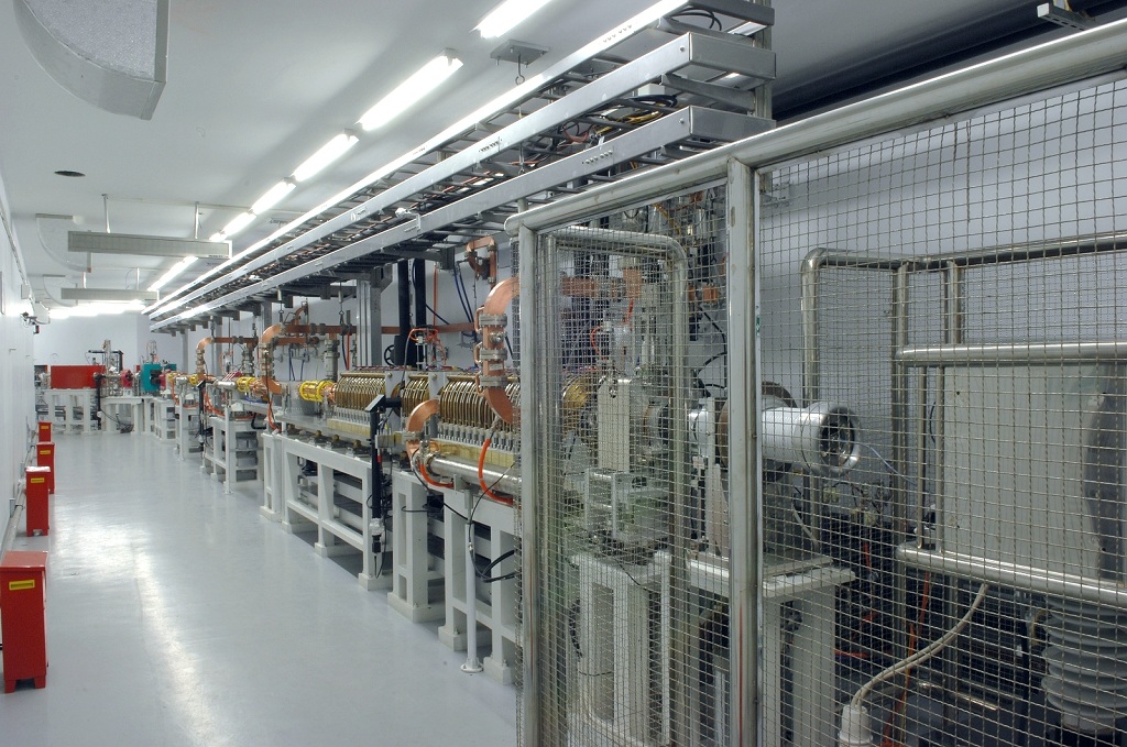
BL14B1 X-ray Diffraction Beamline
The X-ray diffraction beamline (BL14B1) is one of the 7 phase I beamlines at Shanghai Synchrotron Radiation Facility. It is based on a bending magnet light source which is dedicated to x-ray diffraction studies. It has three critical components:a collimating mirror (Rh coated on Si), a sagittally focused double crystal monochromator and a focusing mirror (Rh coated on Si), which can further focus the beam to a size of 0.5 mm x 0.5 mm. The X-ray energy is from 4 ~ 22 keV.
X-ray diffraction (XRD) is the coherent scattering of X-rays by atoms in the lattice. As a traditional experimental method, XRD has the strong vitality and broad applications in the frontier areas such as superconducting materials, catalyst materials, structure determination of three-dimensional biological macromolecules, drug and polymer, nano-materials, surface and interface, semiconductor superlattice, defects. BL14B1 focuses on material science, condensed matter physics fields, designs to investigate powder, -

BL14W1 X-ray Absorption Fine Structure Spectroscopy (XAFS) Beamline
Beamline BL14W1 specializes in x-ray absorption fine structure (XAFS). It has been open for users since May 2009. It is tunable by two types of double crystal monochromator for covering photon energy from 4.5 keV to 50 keV. K-edge absorption of Titanium up to Lanthanum can be studies. Other heavier atomic species can be investigated via L edges. The bulk of our scientific program is in catalysis, materials science and environmental research.
-

BL15U1 Hard X-ray Micro-Focusing Beamline
BL15U1 is a multitechnique hard X-ray microprobe beamline for the energy range 5-20 keV, combined with micro-X-ray fluorescence (μ-XRF), micro-X-ray absorption (μ-XAS), and micro-X-ray diffraction (μ-XRD). The beamline has opened to user since May, 2009, and it’s being applied for a variety of scientific topics such as materials sciences, life sciences, earth and environmental sciences, and archaeology. Hard x-ray microprobe using K-B mirrors is in operation since opening, with a spatial resolution ~2 μm. A nano-focusing system by using zone-plate is developed also,with a beam size smaller than 200 nm at 10keV.
-

BL16B1 Small Angle X-ray Scattering (SAXS) Beamline
The beamline (BL16B1) is primarily a combined small-angle and wide-angle scattering instrument. Small angle and wide angle X-ray scattering (SAXS and WAXS), when coupled with synchrotron radiation, offers research opportunities in studying the microstructure, kinetics, dynamics and rheology of complex and nanostructured materials (polymers, colloids, fluids, liquid crystals, etc) over a wide range of length (e.g., from Angstroms to microns) and time (e.g., from microseconds to hours) scales. For example, the changes in nano-, micro- and supra-molecular structures of biological and synthetic macromolecules, including ionomers, block copolymers, and dendrimers, in solution and in melts, as well as gels and colloidal systems; the kinetics of phase transition in semi-crystalline polymers, blends and nanocomposites.
-

Hard X-ray Spectroscopy Beamline
The Hard X-ray Spectroscopy Beamline (BL11B) is a hard X-ray beamline dedicated to X-ray absorption spectroscopy. BL11B can provide an energy range of 5~30 keV and a focused beam of ~ 250×250 μm2. The main experimental methods include X-ray absorption fine structure (XAFS), quick-scanning XAFS (QXAFS) for time resolved XAFS (ms~s) and combination of XAFS and XRD techniques. The beamline is equipped with highly sensitive 36-element Ge solid state detector (Canberra, for fluorescence XAFS) and one-dimensional MYTHEN 5K detector (for XRD). At present, various in situ experimental devices have been developed, including electrochemical reaction cells (transmission and fluorescence XAFS, XRD), closed-cycle cryostat (~3 K,Montana S50), high temperature and high pressure cells (500℃, 10 bar), gas chromatography-mass spectrometry (GC-MS). Based on the multiple experimental methods, users can carry out frontier scientific research in the fields of physics, energy science, materials science and environmental science, etc. -

BL17U1 Macromolecular Crystallography Beamline
High resolution and high throughput structural determination of macromolecules and their complexes:
l Large macromolecular assemblies
l Membrane proteins
l Structural genomics and Structure-based drug design -
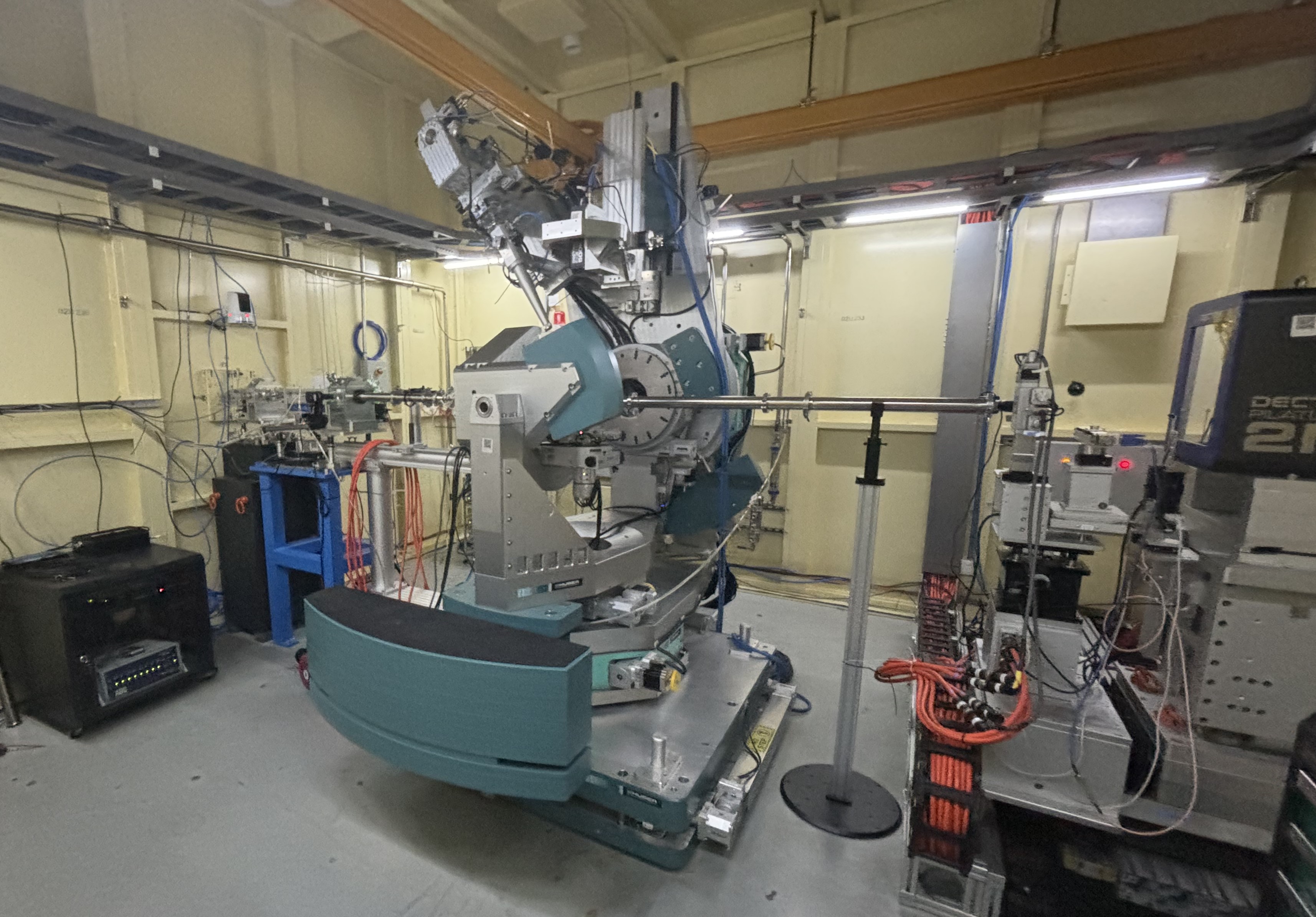
Surface diffraction beamline
Surface diffraction beamline is a high-resolution X-ray diffraction beamline for investigating the structure of surfaces and interfaces of low-dimensional materials, which has the advantage of high brightness, high flux, high energy resolution, and low divergence. This beamline aims to study the surface (solid-vacuum, solid-gas) and interface (solid-solid, solid-liquid, and liquid-liquid) in nanoscience, condensed matter and soft matter systems by mean of various surface scattering techniques. Following highlight research fields will be mainly covered in this beamline: surface and interface of low-dimensional thin films, atomic-level structure of solid surfaces, solid-liquid, liquid-liquid interfaces, biomembrane structure, self-assembly in soft matter. -
BL05U-D-line EDXAS endstation
D-line ED-XAS endstation can provide time-resolved ED-XAS experiments and ED-XAS experiments at extreme conditions (50 GPa, ~3000 K, ~20 T, etc.). This technique has an important applications in the research frontiers such as catalysis, condensed matter, high-pressure physics, nanomaterials, and so on. -

Infrared Spectromicroscopy Station
The spectral range of infrared (IR) spectromicroscopy station (BL06B) covers the near, middle and far infrared region (10cm-1-10000cm-1) at Shanghai Synchrotron Radiation (SSRF). The endstation is equipped with high-resolution Fourier Transform Infrared (FTIR) spectrometer, IR microscope, scanning near-field optical microscope, high-sensitivity MCT detector and various in-situ experimental devices. The main experimental methods include IR spectroscopy, IR spectromicroscopy, nano-resolution IR spectroscopy, and the combination of IR and ED-XAS, etc. SRIR source has the advantages of small light source size, high collimation and high brightness. The diffraction-limited spatial resolution can be obtained by SRIR spectromicroscopy technology. SRIR technology is able to get some microscopic information which is difficult to get by conventional IR source. SRIR technology is widely used in life science, chemistry and polymer science, earth and space science, cultural heritage and archaeology, etc.
-
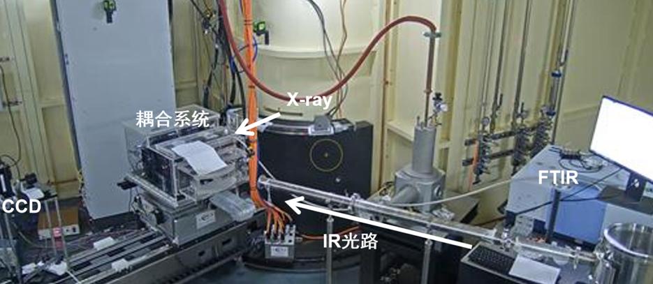
BL05U&06B-D-line EDXAS& IR endstation
Dynamic line (D-line) at SSRF is a beamline which can perform ED-XAS and SR-IR experiments simultaneously at the same sample position. Simultaneous probes of ED-XAS and IR can provide multi-scale (atomic, electronic and molecular) structural information, which can help to better understand the non-equilibrium phenomena. This combining technique has important applications in the field of catalytic reaction and catalytic kinetics. -

Spatial and Spin resolution ARPES and Magnetism
he Spatial and Spin beamline (S2-line) is a soft X-ray beamline to investigate the spatial, spin, energy and momentum information of electron structure. Two APPLE-II type undulators are utilized to provide photons of different polarization states in the energy range of 50-2000eV. The beamline has two branches: one dedicated to Nano angle resolved photoemission spectroscopy (Nano ARPES) that has spatial resolution with 200 nm in level, another branch has three endstations: Spin ARPES has the ability to analyze electron spin; Vector field Station can provide magnetic fields in any directions; High field Station can provide extreme cold temperature and strong magnetic fields.
-
Time-resolved USAXS beamline
Time-resolved USAXS beamline is a bemline dedicated to study microstructures of materials by hard X-ray ultrasmall angle scattering from nanoscale to microscale. The energy of the beamline covers from 8keV to 15KeV. At the beamline, the experimental methods contain time-resolved ultra-small angle X-ray scattering (USAXS), and small angle X-ray scattering (SAXS)/ wide angle X-ray scattering (WAXS) techniques, microfocus SAXS technology, etc.. The scientific goals of the beamline focus on: 1) the internal multi-level structural evolution mechanism of materials in the field environment or in the industrial processes; 2) structural evolution during self-assembly and non-equilibrium dynamics; 3) local microstructures and structural evolution of the materials.
There are three endstations: USAXS endstation, microfocus SAXS endstation and industrial application endstation. The photon flux at the USAXS focus point is about 1.0×10¹³phs/s. The focus beamsize is about 10μm×10μm at microfocus endstation. At microfocus SAXS endstation, microfocus small-angle X-ray scattering technique is avaible to analyze the microdomain structure of materials; at industrial application endstation, time-resolved small-angle/wide-angle X-ray scattering is used to study structural evolution behavior and mechanism of polymer and fiber during processing processes. -

BSL-2 Macromolecular Crystallography Beamline
BSL-2 Macromolecular Crystallography Beamline (BL10U2) is one of the 16 beam lines constructed in the SSRF Phase-II project. This beamline is designed as a crystal diffraction data collection laboratory with a biosafety level-2 protection, in which virus structure and function research by crystal diffraction method is conducted. This beamline will support researchers to study the structure and function of the overall particles of pathogenic microorganisms (such as viruses, etc.) and transcriptional replication complexes, as well as clarify the molecular mechanism of pathogenic microorganisms infecting host cells and transcriptional replication. This beamline will strongly push the development of new, efficient and specific detection techniques, vaccines and therapeutic drugs.
NOTICE
-
Call for Proposals for HEPS Phase II Beamlines May 23,2022